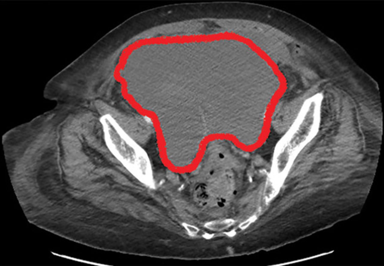
Paracolic Gutter Hematoma

Laparoscopic View Of Left Paracolic Gutter Hematoma Download Scientific Diagram
A Complication Of Enoxaparin Injection Cleveland Clinic Journal Of Medicine

Subcapsular Haematoma Of The Spleen Complicating Acute Pancreatitis Bmj Case Reports
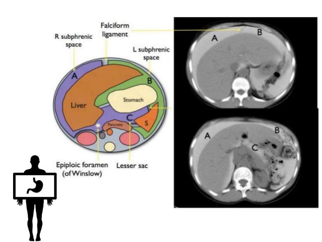
Peritoneum Intraperitoneal Spaces

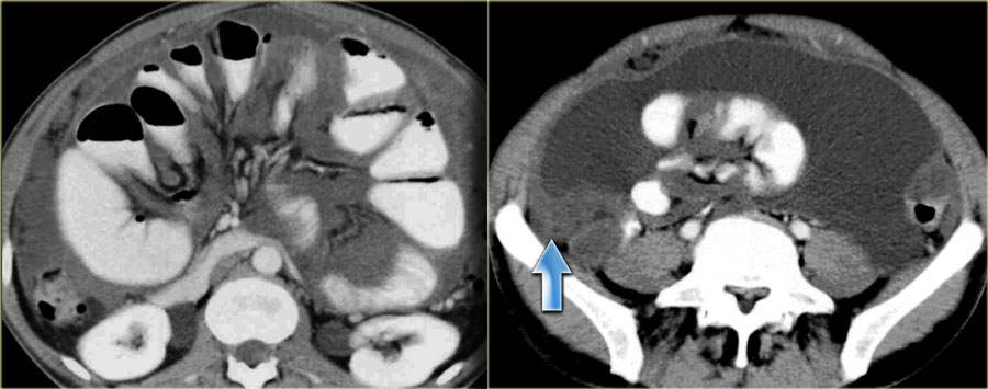
Ultrasonogram Revealed Free Fluid In The Paracolic Gutter Right And Download Scientific Diagram

Amicus Illustration Of Amicus Injury Abdominal Abdomen Intraperitoneal Fluid Pericolic Gutter Dilation Small Bowel Side Wall Pelvic Hematoma Increased Blood Extraperitoneal Spaces Retrovessical Pouch Bladder Compressed Perisplenic Fracture Laverations
Postoperative computed tomography scan of the abdomen showing extensive hemoperitoneum below the spleen in the left paracolic gutter in front of urinary bladder measuring 12 cm in dimension and a distended rectum measuring 8 5 5.
Paracolic gutter hematoma.

Ultrasonogram Revealed Free Fluid In The Paracolic Gutter Right And Download Scientific Diagram
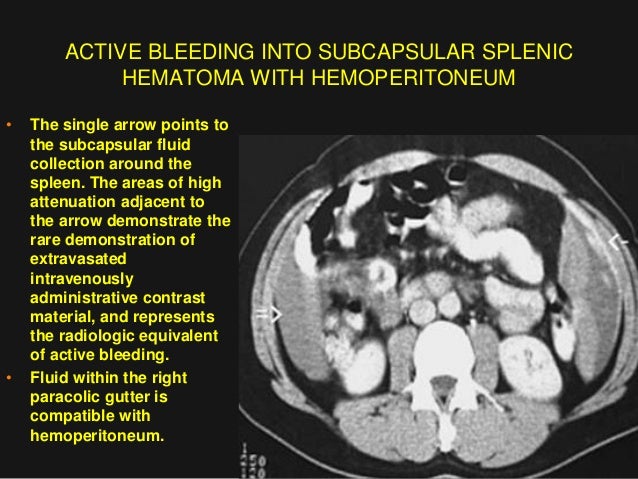
Imaging Abdomen Trauma Spleenic Trauma Part 3 Dr Ahmed Esawy

Ultrasound Image Of A Hemoperitoneum Star In The Right Paracolic Download Scientific Diagram
The Radiology Assistant Peritoneal Pathology
Source : pinterest.com
